THE CARDIOVASCULAR SYSTEM: THE BLOOD
I. Introduction
A. The internal environment is regulated by the fluids which bathe the cells:
1. Interstitial fluid (IF) is found outside cells and in between the cells
2. Lymph inside lymph vessels
3. Blood within blood vessels
B. Hematology is the science which studies blood and blood disorders
II. Functions of Blood
A. Blood is critical in the transportation of oxygen, carbon dioxide, nutrients, heat, wastes, and hormones
B. It assists in the regulation of pH, body temperature, and water content (osmotic status) of cells
C. It has a vital role in prevention of blood loss through the various processes known as hemostasis
D. It provides defense mechanisms against pathogens via white blood cells
III. Physical Characteristics of Blood
A. Greater viscosity than water (Water’s viscosity = 1; Blood’s viscosity = 3.0 -4.5)
B. Temperature of blood: 38°C (100.4oF)
C. pH of 7.35 to 7.45 (Below 7.35 = Acidosis; Above 7.45 = Alkalosis)
D. Blood constitutes approximately 8% of body weight
E. Blood volume ranges between 4 to 6 liters (for females: 4-5 L; for males: 5-6 L)
IV. Blood components
A. 55% plasma and 45% formed elements (Hematocrit “crit” for males: 40%-54% (47%); Females: 38%-46% (42%)
B. Blood plasma composition
1. 91.5% water
2. 8.5% solutes
a. Proteins (albumins; globulins (alpha, beta, gamma); fibrinogen)
b. Nutrients
c. Enzymes
d. Hormones
e. Respiratory gases (oxygen, carbon dioxide)
f. Electrolytes (sodium, chloride, potassium)
g. Waste products (urea, uric acid, creatinine, H+, etc)
C. Formed Elements
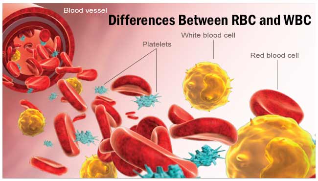
1. Erythrocytes (Red Blood Cells or RBC’s)
2. Leukocytes (White Blood Cells or WBC’s)
3. Thrombocytes (platelets) ,
D. Hematocrit represents the percentage of red blood cells in blood (called Packed Cell Volume (PCV) in animals
1. A lower than normal hematocrit is representative of a condition known as anemia
2. An abnormally high hematocrit is representative of polycythemia
V. Formation of Blood Cells
A. Blood cells are formed from pluripotent hyematopoietic stem cells found in the red bone marrow (myeloid)
1. Myeloid stem cells give rise to RBC’s, platelets, and all WBC’s except lymphocytes
2. Lymphoid stem cells give rise to lymphocytes
B. Hemopoiesis (hematopoiesis) is stimulated by several hematopoietic growth factors. These hematopoietic
growth factors stimulate differentiation and proliferation of blood cells
1. Erythropoietin increases the number of RBC precursors
2. Thrombopoietin increases the number of platelet precursors
3. Cytokines (colony stimulating factors and interleukins) increase the number of WBC precursors
VI. Red blood cells
A. Erythrocytes (Red Blood Cells) contain the oxygen-carrying protein hemoglobin
B. There are approximately 5.4 million red blood cells/microliter of blood (1 drop of blood = 50uL)
C. Erythrocytes are biconcave discs without nuclei
D. RBC physiology
1. Hemoglobin functions to transport oxygen (oxyhemoglobin) and carbon dioxide (carbaminohemoglobin)
2. Hemoglobin functions to regulate blood pressure
a. Unsaturated hemoglobin picks up CO2 and nitric oxide (NO), a gas which it releases to
the lungs to be exhaled
b. Saturated hemoglobin carries fresh oxygen and supernitric oxide (SNO) from the lungs
and releases them in body tissues.
c. NO tends to cause vasoconstriction; SNO causes vasodilation
d. By ferrying NO and SNP throughout the body, hemoglobinhelps regulate blood pressure
by adjusting the amount of NO or SNO to which blood vessels are exposed.
E. RBC Life Cycle
1. Red Blood Cells only live about 120 days because of the wear and tear on their plasma
membranes as they squeeze through blood capillaries
2. In the RBC life cycle, after phagocytosis of warn-out RBC's by macrophages, hemoglobin is
recycled; the globin portion is split from the heme with the amino acids being reused for protein
synthesis. The iron in the heme portion is reclaimed with the rest of the heme molecule; the rest
becomes a component of bile in the digestive process.
F. Erythropoiesis (production of RBC's)
1. Erythropoiesis is the production of red blood cells in the red bone marrow of some bones
2. Proerythroblasts give rise to cells which begin to synthesize hemoglobin
3. At the end of development, the nucleus is ejected (causing it to become biconcave) and becomes reticulocyte
4. Reticulocytes pass from the bone marrow to the circulatory system; now are called erythrocytes
5. Hypoxia (low cellular oxygen) is detected by cells within the kidney which stimulates the
production of erythropoietin
6. Erythropoietin travels to the red bone marrow where it speeds the development of pro-erythrocytes
into reticulocytes
Vll. White blood cells
A. Leukocytes ( WBC’s) are nucleated cells, do not contain hemoglobin, but do contain cytoplasmic granules.
1. Granular leukocytes include eosinophils, basophils, and neutrophils based on the staning of
the enclosed granules.
2, Agranular leukocytes have small, resistant-to-staining-cytoplasmic granules and included lymphocytes
and monocytes (which differentiate into fixed and wandering macrophages)
B. Leukocytes have surface proteins, as do erythrocytes. Thse are called major histocompatibility antigens
(MHC), are unique for each person (except for identical siblings), and can be used to identify a tissue.
C. WBC physiology
1. White blood cells usually live for only a few hours or a few days
2. Normal blood contains 5,000 to 10,000 leukocytes/uL
3. Leukocytosis refers to an increase in the number of WBC's (-philia for increased eos, basos, neuts)
4. Leukopenia refers to an abnormally low number ofWBC's (-penia for decrease in any WBC)
5. The general function of leukocytes is to combat inflammation and infection
a. WBC's leave the blood stream by emigration.
b. Some WBC’s particularly neutrophils and macrophages, are active in phagocytosis
c. Chemotaxis is the chemical attraction of WBC’s to a disease or injury site
d. Different WBC’s combat inflammation and infection in different ways:
(1) Neutrophils and wandering or fixed macrophages (which develop from monocytes
do so through phagocytosis
(2) Eosinophils combat the effects of histamine in allergic reactions, phagocytize, antigen-
antibody complexes, and combat parasitic worms
(3) Basophils develop into mast cells that liberate heparin, histamine, and serotonin in
allergic reactions that inteniify the inflammatory response
(4) B-lymphocytes, in response to the presence of foreign substances called antigens,
differentiate into plasma cells that produce antibodies (humoral-mediated immunity) and
T-lymphocytes destroy foreign invaders directly (cell-mediated immunity)
NK lymphocytes kill a wide variety of infectious microbes and certain self-tumor cells.
(5) Monocytes emigrate from blood to tissues where they enlarge and differentiate into either fixed
or wandering macrophages; depending on where they reside are called Langerhan’s cells (skin),
Kupffer’s cells (liver sinusoids), alveolar cells (lung); osteoclasts (bone c.t.), etc.
e. A differential white blood cell count is a diagnostic test in which specific white blood cells are
enumerated. Because each type of WBC plays a different role, determining the percentage of each
type in the blood assists in diagnosing the condition
6. Bone marrow transplants may be used to treat several types of anemia, leukemia, and other blood disorders
VIII. Platelets
A. Thrombopoietin stimulates myeloid stem cells to produce platelets
1. Myeloid stem cells develop into megakaryocyte-colony-forming cells that develop into megakaryoblasts
2. Megakaryoblasts transform into megakaryocytes, which ultimately fragment
3. Each fragment, enclosed by a piece of cell membrane, is a platelet (thrombocyte)
B. Normal blood contains 250,000 to 400,000 platelets/uL.
C. Platelets have a life span of only 5 to 9 days. Aged and dead platelets are removed by fixed macrophages in the spleen and liver
D. Platelets help stop blood loss from damaged vessels by forming a platelet plug. Their granules also
contain chemicals that promote blood clotting.
IX. Hemostasis
A. Hemostasis refers to the stoppage of bleeding. When blood vessels are damaged or ruptured,
the hemostatic response must be quick, localized to the region of damage, and carefully controlled.
B. Hemostasis involves vascular spasm, platelet plug formation, and blood coagulation (clotting)
1. Vascular spasm--the smooth muscle of a vessel wall will contract to constrict vessel’s lumen
2. Platelet plug formation--the clumping of platelets around the damage to slow bleeding
3. A clot is a gel consisting of a network of insoluble protein fibers (fibrin) in which formed
elements of blood are trapped
a. The chemicals involved in clotting are known as coagulation (clotting) factors; most are in blood plasma;
some are released by platelets, and one is released from damaged tissue cells
b. Blood clotting involves a cascade of reactions that may be divided into three stages:
(1) De novo formation of prothrombinase (prothrombin activator)
(2) Conversion of prothrombin (circulating clotting protein) into thrombin
(3) Conversion of soluble fibrinogen into insoluble fibrin threads.
C. Normal coagulation is a positive feedback mechanism; requires Vitamin K; also involves clot retraction
(tightening of the clot) and fibrinolysis (dissolution of the clot)
1. The fibrinolytic system dissolves small, inappropriate clots and clots at a site of damage once
the damage is repaired
2. Plasmin (fibrinolysin) can dissolve a clot by digesting fibrin threads and inactivating
substances such as fibrinogen, protrhombin, and factors V, VIII, and XII.
D. Homeostatic Control of Hemostasis
1. Clots are generally localized due to fibrin absorbing thrombin into the clot, clotting factors
diffusing through blood, and the production of prostacyclin, a powerful inhibitor of platelet adhesion
and release
2. Substances that inhibit coagulation, called anticoagulants (such as heparin) are also present in blood.
E. Despite the anticoagulating and fibrinolytic mechanisms, blood clots sometimes form within the
cardiovascular system
1. Clotting in an unbroken blood vessel is called thrombosis; the clot is called a thrombus.
2. A thrombus (clot), bubble of air, fat from broken bones, or piece of debris transported by
the bloodstream that moves from the site of origin is called an embolus.
X. Blood Groups and Blood Types
A. The surfaces of red blood cells contain genetically determined blood group antigens, called
agglutinogens or isoantigens.
1. Blood is categorized into different blood groups based on the presence or absence of various isoantigens.
2. Within a blood group there may be two or more different blood types.
3. Major blood groups are the ABO and Rh groups. Other blood groups include the Lewis, Kell, Kidd, and Duffy systems.
B. ABO Group
1. In the ABO system, agglutinogens (antigens) A and B determine blood type.
2. Plasma contains agglutinins (antibodies), designated as a and b, that react with